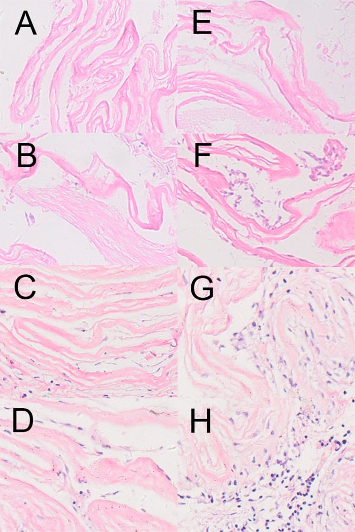
Figure 3.
HE staining of the cell-seeded dHAS and dHAS after subcutaneous implantation. (A–D): HE stainings of cell-seeded dHAS engrafts at week 1, 2, 4, and 8 after subcutaneous implantation; (E–H): HE stainings of dHAS engrafts at week 1, 2, 4, and 8. Fewer inflammatory infiltrations in dHAS xenografts were found, in comparison to AM grafts (magnification at ×200).

