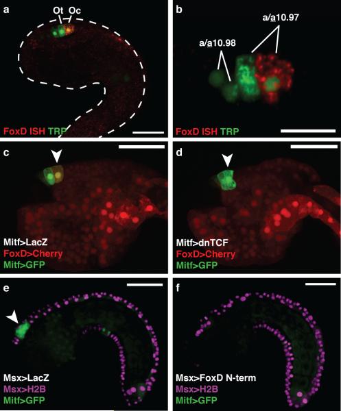
Figure 2. FoxD represses Mitf in the ocellus.
a, Tailbud electroporated with TRP>LacZ detected with an antibody (green) marking the precursors of the otolith and ocellus, and hybridized with a FoxD probe (red). b, FoxD is expressed in the posterior a10.97 cell. c-d, Tailbuds electroporated with FoxD>mCherry and Mitf>GFP. Arrowheads mark the presumptive ocellus. c, Co-electroporated with Mitf>LacZ (126/180 expressed mCherry). d, Co-electroporated with Mitf>dnTCF (only 30/180 expressed mCherry). e-f, Tailbuds electroporated with Msx>H2B mCherry, and Mitf>GFP. Arrowhead shows GFP expression in a9.49 derivatives. e, Co-electroporated with Msx>LacZ. f, Co-electroporated with Msx>FoxD N-term. Scale bars 50 μm (a, c-f); 25 μm (b).

