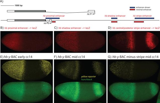
Fig. 1. Summary of Hb Cis-Regulatory DNAs.
(A) The Hb locus contains two promoters, P2 and P1, and three enhancers. The proximal and distal shadow enhancers are targets of the Bicoid gradient, while the stripe enhancer is regulated by gap repressors (see text). (B-D) lacZ antisense RNA in situ hybridization assays with transgenic embryos expressing proximal->lacZ (B), shadow->lacZ (C), or stripe->lacZ (D) transgenes. The proximal and shadow enhancers mediate broad expression in anterior regions, while the stripe enhancer produces central and posterior stripes of expression. (E-G) Transgenic embryos expressing a y-BAC transgene containing a 44 kb genomic DNA encompassing the Hb locus and associated regulatory DNAs. The Hb transcription unit was replaced with the yellow (y) reporter. The wild-type y-BAC transgene exhibits broad anterior expression and a posterior stripe (E,F), whereas a mutagenized y-BAC transgene containing an internal replacement of stripe enhancer sequences with a non-regulatory spacer exhibits reduced expression of the central and posterior stripes (G). The embryos were double stained for yellow nascent transcripts (y-BAC transgenes) (shown in yellow) and endogenous hb (shown in green).

