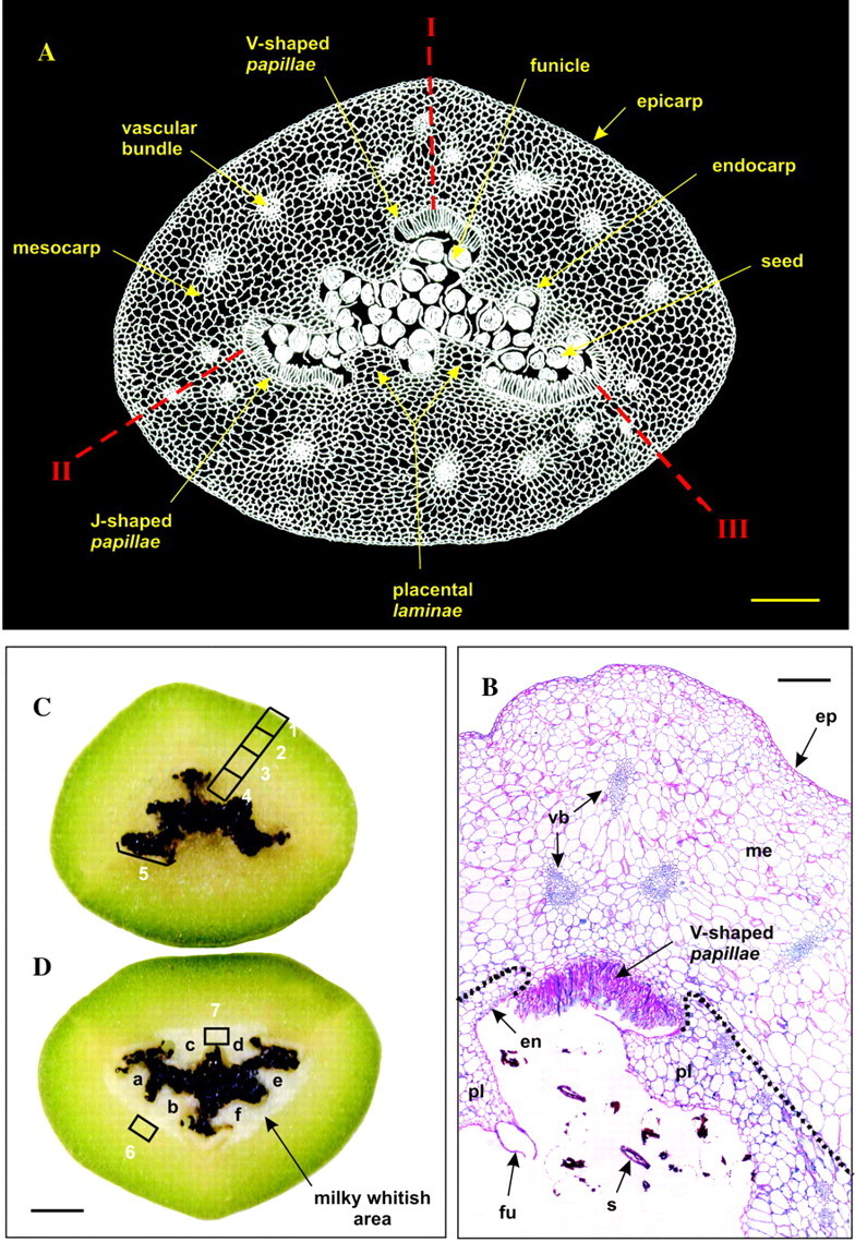
Fig. 1. General views of a transverse equatorial section of a mature green vanilla bean. A, Cross-section drawing of a mature vanilla bean (negative image). Notice the carpel suture lines (I, II, III) and the longitudinal dehiscence splits (II, III). B, Thin cross-section (5 µm thick) of a mature vanilla bean after staining with periodic acid–Schiff (PAS)–Naphthol Blue Black. ep, Epicarp; me, mesocarp; en, endocarp; pl, placental lamina; fu, funicle; s, seed remnants; vb, vascular bundle. The dotted line represents the boundary demarcating the milky whitish area visible in D. C, Cross-section of a fresh, mature green vanilla bean. D, Cross-section of a frozen, mature green vanilla bean. In C and D, zones 1–7 were hand-dissected and analysed for glucovanillin and β-glucosidase contents. Notice that freezing led to the appearance of a frosted, milky whitish area clearly separated from the outer part of the bean by a sharp boundary. a–f, Placental laminae. Bars = 0·15 cm (A), 400 µm (B) and 0·3 cm (C, D).
