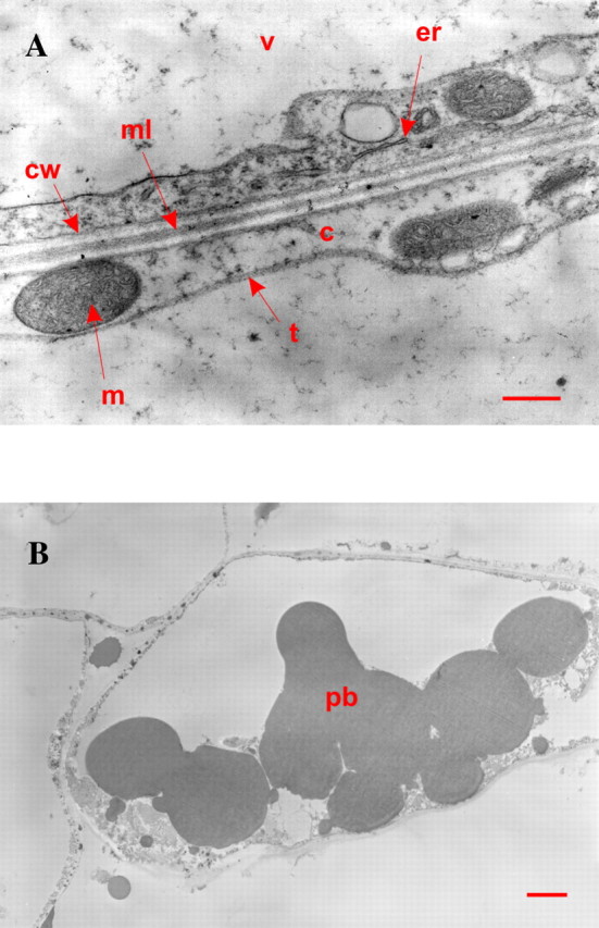
Fig. 3. Transmission electron micrographs of cells from the placental laminae. A, View of a cell edge. c, Cytoplasm; m, mitochondrion; er, endoplasmic reticulum; v, vacuole; t, tonoplast; cw, cell wall; ml, middle lamella. Bar = 500 nm. B, View of a cell exhibiting aleurone protein bodies. Bar = 2 µm.
