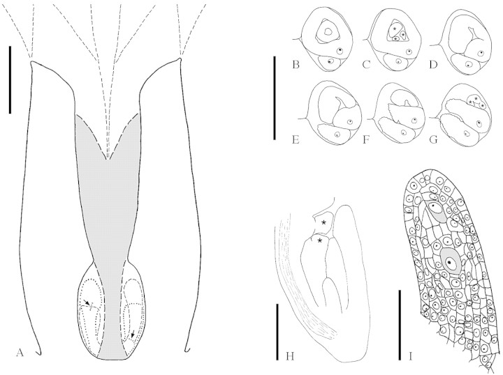
Fig. 7.Balanops vieillardii. A, Schematic median LS of gynoecium at anthesis (upper stigmatic region not drawn). Post-genitally fused area is shaded. Outline of parts that are not exactly in the median plane interrupted. Micropyles are marked with arrows (longitudinal in longer ovule, transverse in shorter ovule). B–G, TS series of the two ovules in a locule, shorter ovule showing micropylar region, longer ovule showing funicle. Vascular bundle in raphe indicated (xylem black). Lobes of inner (in C) and outer (in G) integument marked with stars. B, Level of both integuments; C, lobes of inner integument; D–G, lobes of outer integument. H, Median LS ovule at megaspore mother cell stage. Vascular bundle in raphe indicated. Lobes of inner and outer integument marked with stars. I, Nucellus of the same ovule, showing two potential megaspore mother cells (shaded). Bars: A = 1 mm; B–G = 0·5 mm; H = 0·2 mm; I = 0·05 mm.

An official website of the United States government
Here's how you know
Official websites use .gov
A
.gov website belongs to an official
government organization in the United States.
Secure .gov websites use HTTPS
A lock (
) or https:// means you've safely
connected to the .gov website. Share sensitive
information only on official, secure websites.
