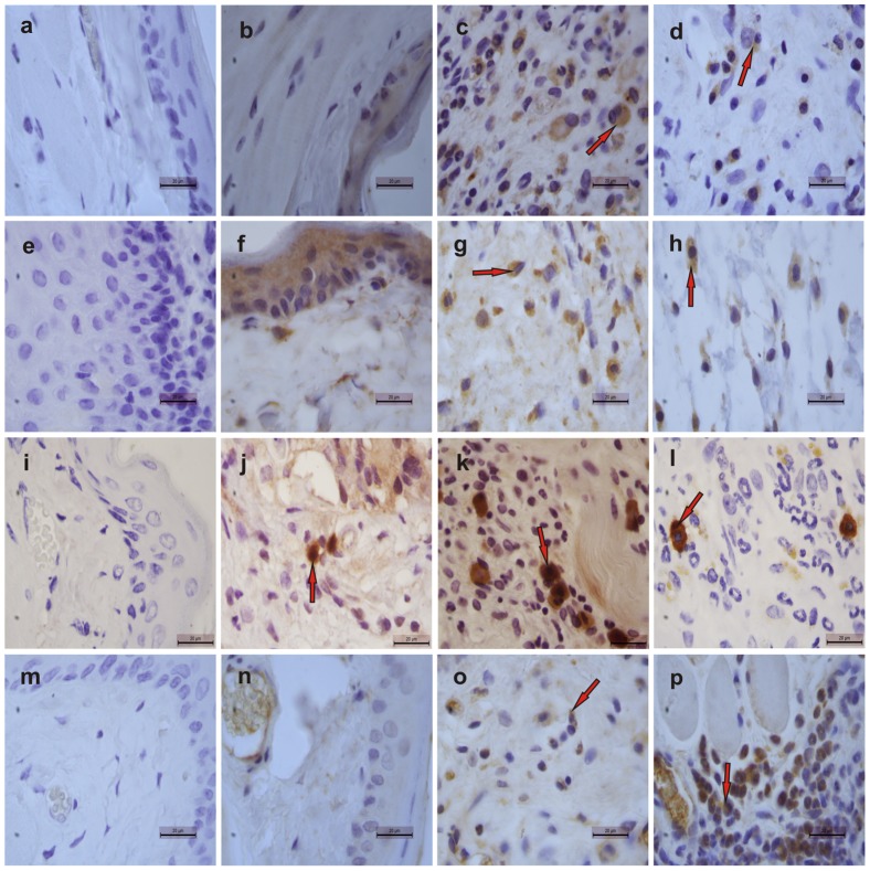
Figure 4. Representative examples of iNOS (1st row), IL-1β (2nd row), TNF-α (3rd row) and TGF-β RII (4th row) immunostaining on day 14 in tissues from cheek pouches of hamsters subjected to 5-FU-induced oral mucositis.
Staining was performed using cheek pouches from healthy animals (b, f, j, n) and animals subjected to 5-FU-induced mucositis that received topical applications of S-nitrosoglutathione (GSNO; 0.5 mM; d, h, l, p) or saline (c, g, k, o). Negative controls were samples of cheek pouches where the primary antibody was replaced with PBS-BSA (5%); no immunostaining was detected (a, e, i, m). Magnification, ×1000.

