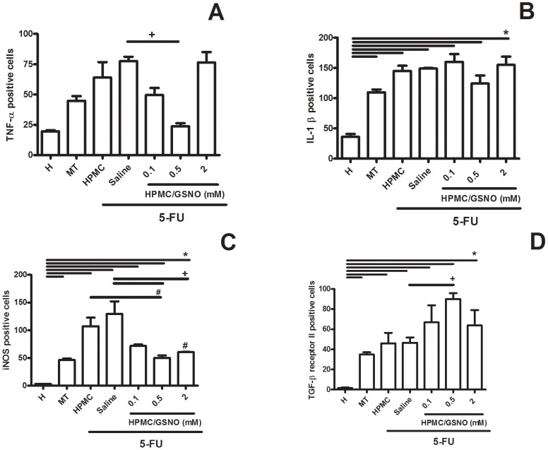
Figure 5. Quantification of TNF-α- (A), IL-1β- (B), iNOS- (C) and TGF-β RII - (D) positive cells in cheek pouch tissues of hamsters subjected to 5-FU-induced oral mucositis, on day 14.
Oral mucositis was induced in hamsters by intraperitoneal (i.p.) injection of 5-FU followed by mechanical trauma (MT) of the cheek pouch. Animals received topical applications of a gel containing 0.5 mM S-nitrosoglutathione (GSNO) 30 min prior to 5-FU and twice daily thereafter for 10 days or 14 days. Control groups comprised healthy animals (H) and animals subjected to 5-FU-induced oral mucositis that received local applications of saline (Saline). Cells positive for staining were counted (10 fields per slide, 400×) for statistical comparisons. Bars denote the means ± standard errors of positive cells from four slides per group (4 animals per group). *denotes significant differences (P<0.05) compared with the Healthy group; +denotes a significant difference (P<0.05) compared with the Saline group; #denotes a significant difference (P<0.05) compared with the HPMC group. Data were analyzed using the Kruskal Wallis and Mann Whitney tests.

