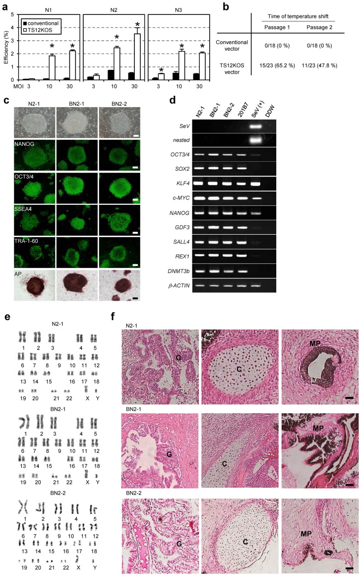
Figure 2. Characterization of human iPSCs generated by the TS12KOS vector.
(a) iPSC generation from human peripheral blood cells. Experiments were conducted in triplicate (mean ± SD). N1, N2, and N3 indicate individual healthy volunteers. *P<0.01, TS12KOS vector versus conventional vectors, Student's t-test. (b) Nested RT-PCR analysis of the elimination of SeV vectors after the temperature shift from 37°C to 38°C. (c) Phase contrast images, immunofluorescence for pluripotency markers, and alkaline phosphatase (AP) staining of iPSC lines. The iPSC lines N2-1 and BN2-1 and BN2-2 were derived from the skin-derived fibroblasts and blood cells of N2 healthy volunteer, respectively. Scale bars, 200 µm. (d) RT-PCR analysis of Sendai virus and human ES cell markers. SeV, first RT-PCR for SeV; nested, nested RT-PCR for SeV; 201B7, control human iPSC line; SeV(+), Day 7 SeV-infected human fibroblasts. (e) Chromosomal analyses of iPSC lines generated with the TS12KOS vector. (f) Tissue morphology of a representative teratoma derived from iPSC lines generated with the TS12KOS vector. G, glandular structure (endoderm); C, cartilage (mesoderm); MP, melanin pigment (ectoderm). Scale bars, 100 µm.

