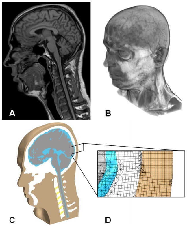
Figure 1. Image-based finite element model of the human head and neck.

(A) Volume rendered image of MRI scan data used to construct this model. (B) Finite element head model subject to an example simulated golf ball impact at head posterior. (C) Isometric section view of model revealing soft and hard tissue structures. (D) Enlarged view of mesh at the occipital region of the head.
