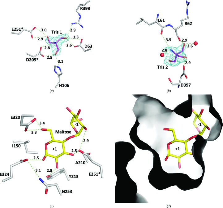
Figure 6.
The substrate-binding site in DrTS. (a) The Tris bound at the −1 subsite in the N253A mutant. The simulated-annealing F o − F c OMIT map for Tris is contoured at the 6σ level. (b) An additional Tris-binding site in the N253A variant. The simulated-annealing F o − F c OMIT map is contoured at the 4σ level. (c) The interaction networks between DrTS and the modelled maltose in the +1 subsite. Hydrogen bonds are shown as green dashed lines. (d) Molecular surfaces of the +1 subsite. The view is the same as that in (c).

