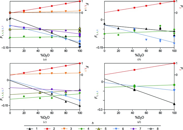
Figure 2.
Structure-factor amplitudes with assigned phases versus %D2O for (a) rat sciatic nerves, (b) rat optic nerves, (c) mouse sciatic nerves and (d) mouse spinal cords. Symbols represent experimental replicates. Lines represent the linear dependence of Fh on %D2O. In the absence of measurements of F 8 in rat sciatic nerve at multiple concentrations of D2O, the linear regression of F 8 versus %D2O was forced through 0 at 100% D2O. Similarly, F 6 for mouse spinal cord was modelled using the single observed reflection at 100% D2O-saline and the slope of F 6 versus %D2O from rat optic nerve. The y-axis labels are colour-coded to correspond to the respective data sets.

