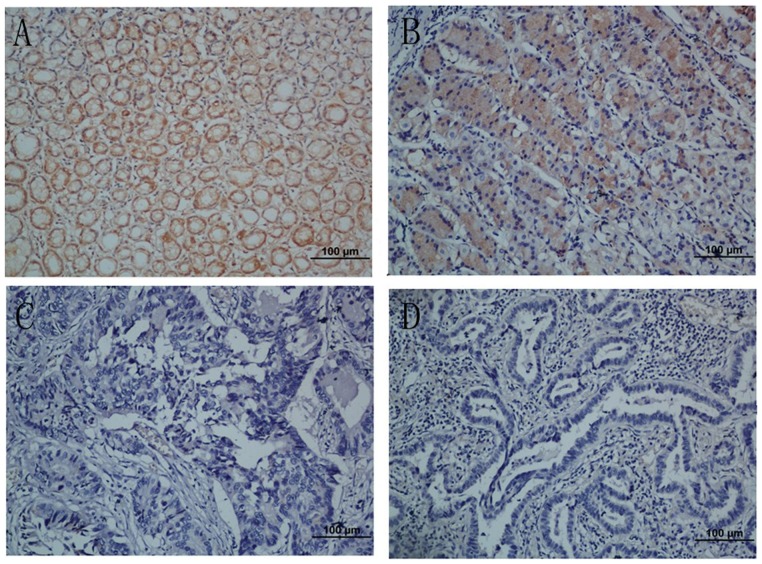
Figure 3. Immunohistochemical analyses of NKX2.1 expression in resection specimens of primary gastric carcinoma.
(A) Strong NKX2.1 staining was observed in noncancerous gastric mucosa glands. (B) Immunostaining of the well-differentiated gastric cancer cells. (C) Weak NKX2.1 staining in poorly-differentiated gastric adenocarcinoma. (D) NKX2.1-negative gastric adenocarcinoma. Original magnification for A–D, ×200.

