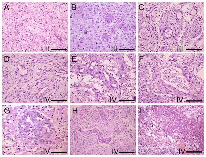
Figure 1. The morphology changes of glioma microvasculature along with the increase of the WHO grade.

(A) thin-wall or sinusoid vessels with different sizes of lumen in grade II glioma, (B-C) irregular buds, cell cords and thick-wall vessels in some areas of grade III glioma, (D-H) strip cord, plexus, glomeruloid, ophidian microvessels in grade IV glioma and (I) more proliferation of heterotypical vessels found around necrotic and hemorrhagic areas in grade IV glioma. (HE A-G Bar = 100 um, H and I Bar = 200 um).
