
Figure 3. Immunohistochemical double staining against CD34 and α-SMA.
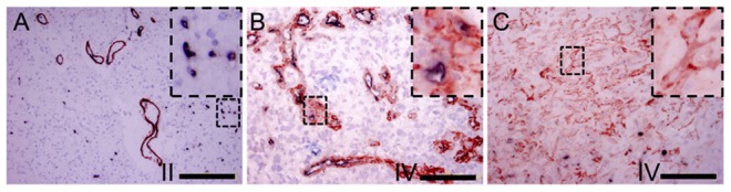
(A) The inner layer of thin microvessels showed CD34 dark purple staining while the closely connected outer layer was in α-SMA red staining in WHO grade II glioma (Bar = 200 um). (B) Multiple layer of microvessels cells were α-SMA positive while only the lining cells of the vessels were CD34 positive. (C) In some areas of WHO grade IV glioma, focal, tubular or cordal cell clusters were found to be α-SMA positive only.(Bar = 100 um).