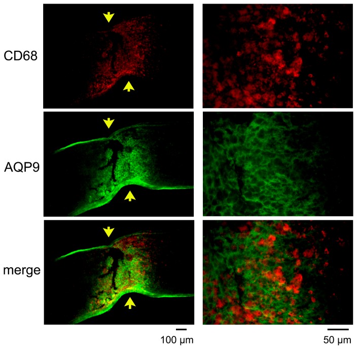
Figure 7. Double labeling of AQP9 and CD68 at the crushed site on day 7.
Representative photographs from 3 independent samples are presented with higher magnifications in the right column. Arrows indicate crush sites. CD68 positive cells are present between the AQP9 positive fibrils suggesting that microglia/macrophages are not the cellular sources for AQP9 after crushing the optic nerve.

