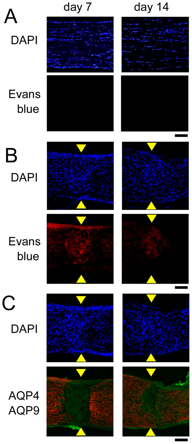
Figure 8. Barrier function determined by extravasated Evans blue.

Evans blue dye has not leaked into the optic nerve tissue in the sham controls (A), but it has leaked at the crushed site by the presence of red fluorescence (B). Merged images of AQP4 (red) and AQP9 (green) indicate that extravasated Evans blue was restricted to the crushed site, where AQP4 was negative but AQP9 was expressed (C). The left and right columns are images on days 7 and 14, respectively. Arrowheads indicate crush sites. Bar = 100 µm.
