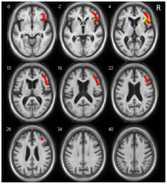
Figure 3. Brain Regions with Relative Hypometabolism (red) and Relative Gray Matter Atrophy (yellow) in Relation with Hallucinations.

Relative hypometabolism of right inferior frontal gyrus, middle frontal gyrus (including orbital part), and insula (anterior part) (slices z = −8 to 28). Core regions correspond to the common part of relative hypometabolism and atrophy, including right inferior frontal gyrus and a part of right anterior insula. (For MRI and FDG PET, p<0.001, uncorrected, minimum cluster size = 25 voxels). Right is right.
