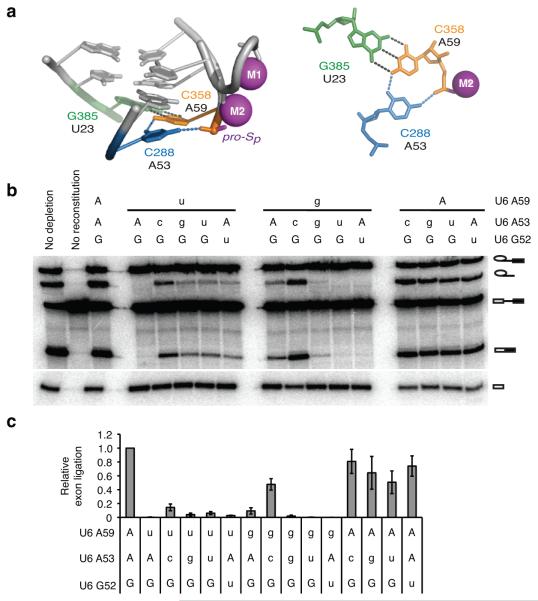
Figure 8. The U6 triplex promotes exon ligation in vitro.
a, Configuration of the C288*C358-G385 base triple in the group II intron (PDB 4FAQ, ref. 8), equivalent to the spliceosomal U6-A53*U6-A59/U2-U23 base triple; left, side view; right, top view. b, Denaturing PAGE analysis of splicing of ACT1 pre-mRNA in extracts reconstituted with the indicated U6 variants. No dep., no depletion; no rec., no reconstitution. Upper case indicates wild-type allele. c, Quantification of exon ligation for the indicated U6 variants, normalized to wild-type U6; exon ligation was calculated as mRNA/lariat intermediate (ref. 19). Error bars represent s.d. of two independent experiments and two technical replicates for each experiment. The efficiency of branching was within 15% of wild type for all U6 variants (quantification not shown). For full gel see Supplementary Fig. 8i.

