
Figure 3.
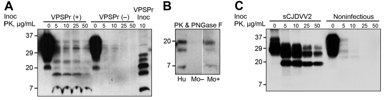
Western blot characteristics of PrPSc recovered from brain of VPSPr-inoculated Tg mice and controls. A) BH treated with increasing amounts of PK show PK-resistant PrPSc fragments with a ladder-like electrophoretic profile in positive VPSPr-inoculated mice, VPSPr (+), even at high concentrations of PK (50 μL/mL). In contrast, nonspecific bands are seen in negative VPSPr-inoculated mice, VPSPr (‒). The banding pattern in VPSPr (+) roughly recapitulates that of the PK-treated PrPSc from the VPSPr inoculum (VPSPr Inoc). B) Positive Tg mice BH treated with PK (25 μg/mL) and PNGase F show 3 PrPSc bands migrating at ≈20 kDa, ≈17 kDa, and ≈7 kDa (Mo +), replicating those of similarly treated VPSPr inoculum (Hu). No bands can be detected in the negative Tg mice (Mo ‒). (All 3 preparations were run on the same gel, but unnecessary lanes were removed). C) Tg mice inoculated with sCJDVV2 BH (from sCJD homozygous valine harboring PrPSc type 2) or inoculated with noninfectious BH used as positive and negative controls exhibit typical PK-resistant PrPSc. Tg(HuPrP129V)×8 BH and monoclonal antibody 1E4 were used in all Western blot tests. Approximate molecular masses are in kDa. BH, brain homogenate; PK, proteinase K; PrPSc, scrapie prion protein; sCJD, sporadic Creutzfeldt-Jakob disease; VPSPr, variably protease-sensitive prionopathy; Tg, transgenic.