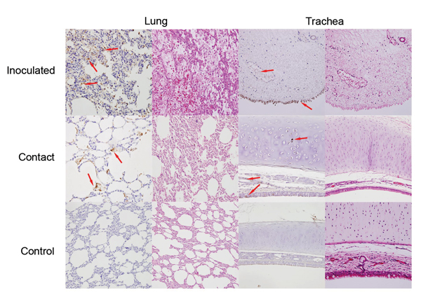
Figure 2.

Detection of viral antigens in the respiratory tract of cats inoculated with equine influenza A(H3N8) virus and from a contact cohort. For each tissue type, the left column shows incubation with a monoclonal antibody against equine influenza virus hemagglutinin and the right column shows hematoxylin and eosin staining. Arrows indicate detection of viral antigen (hemagglutinin) expression (brownish staining). Original magnification ×100.
