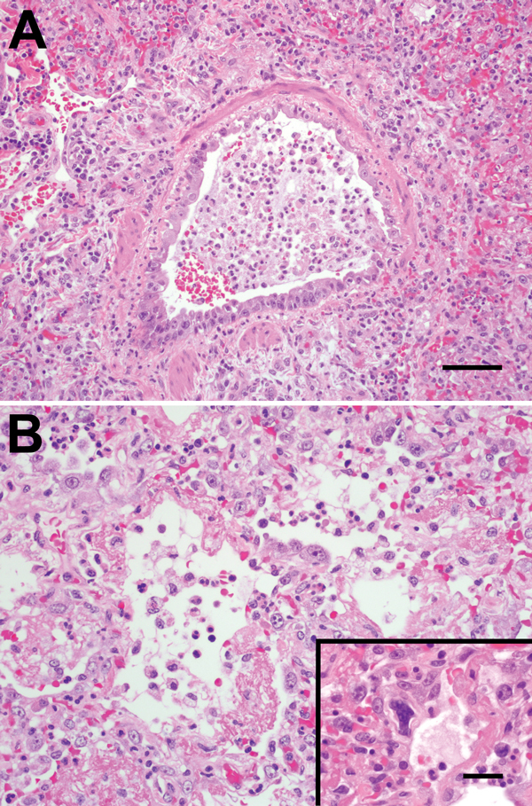
Figure 2.

A) Bronchiolar epithelium of chimpanzees infected with human metapneumovirus, United States, 2009, showing cell variation from attenuated to piled and disorganized. Epithelial cells lack cilia, and lumens contain foamy macrophages, neutrophils, and hemorrhage. Adjacent air spaces are filled with similar inflammatory cells. Scale bar = 70 μm. B) Alveoli lined with plump type II pneumocytes and fibrin. Inset: Rare, deeply basophilic, smudged nuclei are present in some areas. Scale bar = 20 μm. Hematoxylin and eosin stain.
