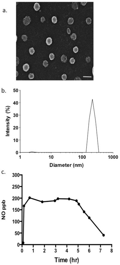
Figure 1.
Characterization of the NO-np. The structure was analyzed using (a) scanning electron microscopy (SEM; bar 100 nm) and (b) analytical sizing performed using dynamic light scattering (DLS). (c)The graph shows the release of NO from the NO-np once placed in an aqueous environment over the course of 8 hours.

