
Figure 4.
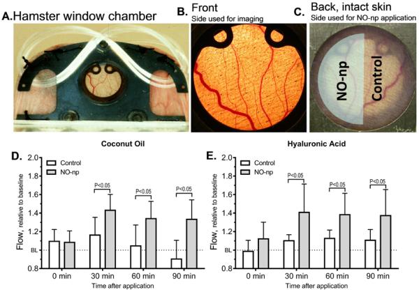
4A, Hamster window chamber model, the front side (4B) was observed using intra vital microscopy and the back side (4C) or intact skin received the NO-np. On one half of the intact skin exposed area the NO-np were applied (shaded) and blood flow compared with the untreated area (control). The control area received the carrier: The difference in microcirculatory blood flow caused by NO-np applied using coconut oil (4D) or hyaluronic acid (4E) as carrier from time of application is shown.