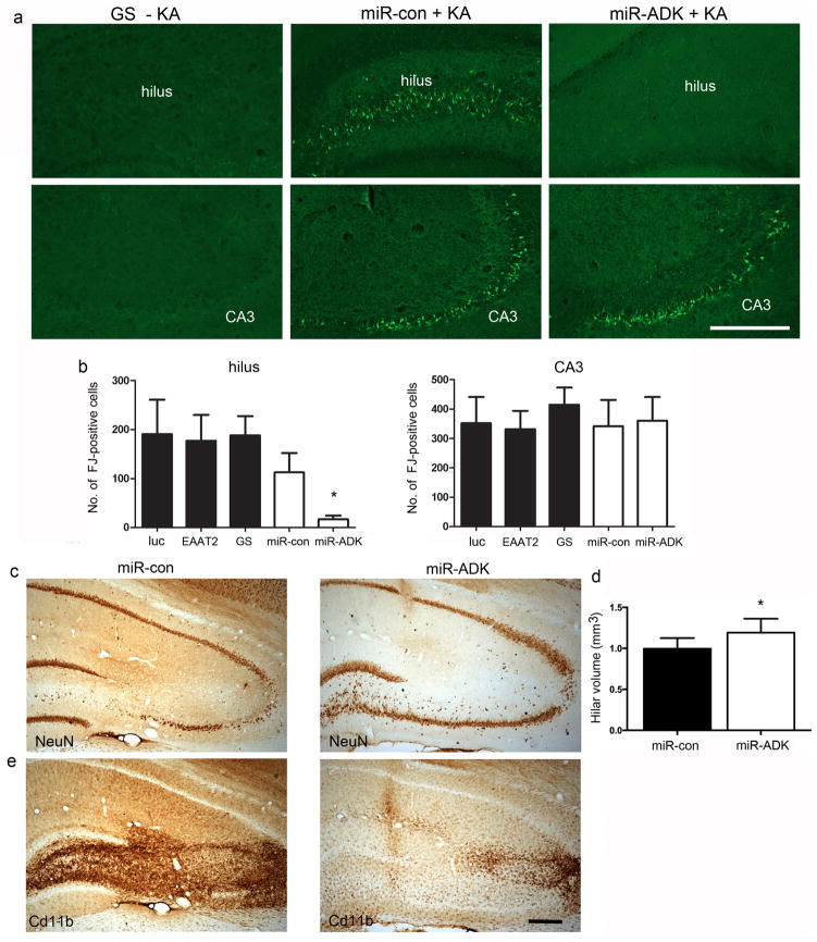
Fig. 4. Attenuated neuronal cell loss in the hippocampus in miR-ADK-treated rats following intrahippocampal kainate.
(a) Representative images of Flurojade-B (FJ) staining in the dentate hilus and CA3 region in brains injected with vector alone (section from GS vector injected brain shown as example (GS –KA)) or miR-con or miR-ADK vector-injected brain 6 days following kainate infusion (miR-con + KA, miR-ADK + KA). (b) Total numbers of FJ-positive cells in the hilus and CA3 region. (c) NeuN immunostaining in the hippocampus and (d) stereological quantification of hilar volumes in NeuN-stained sections in the miR-con and miR-ADK treated rats. (d) Reduced microglial proliferation as visualized by Cd11b-immunoreactivity. For all graphs, bars represent mean + SEM, n = 8–10, Unpaired t-test, *P = 0.02. Scale bar, 300 um

