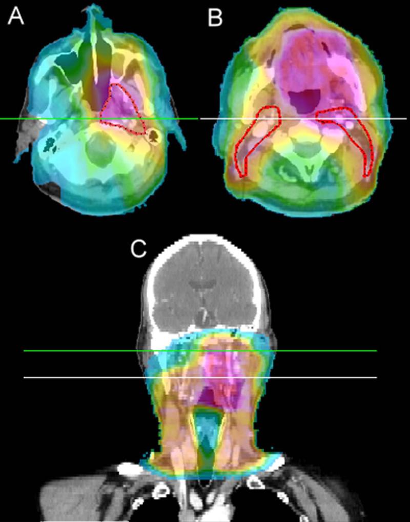
Figure 2.
Dose colorwash CT images of a patient with a T4AN0 oropharyngeal tumor treated in generation 3 which spared the contralateral retropharyngeal lymph nodes and the contralateral high level II lymph nodes. Colorwash range is from 5 Gy to 80 Gy. (A) Axial representation above the spared high level two with the elective CTV2 (56 Gy) volume shown in red. (B) Axial representation below the crossing of the digastric muscle and internal jugular vein also shows the elective CTV2 volume. (C) Coronal depiction shows the spared regions and indicates the axial slice levels.

