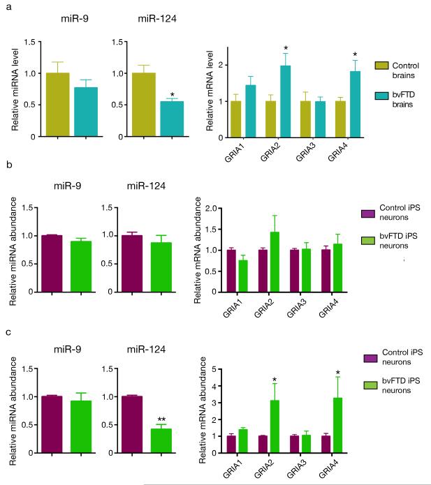
Figure 5.
miR-124 and AMPAR levels are altered in the frontal cortex of subjects with bvFTD and in cortical neurons derived from iPSC lines of three subjects with bvFTD. (a) Quantification of the abundance of miR-124, miR-9, and AMPAR transcripts by RT-PCR in the frontal cortex of subjects with bvFTD and controls. We normalized the content of AMPAR mRNAs against the geometric mean of four different neuronal-specific reference genes (MAP2, Enolase 2, GAP43, and PSD95). *: P < 0.05 by Mann-Whitney test. (b) Quantification of miR-124, miR-9, and AMPAR transcripts in 2-week old human neurons derived from 3 iPSC lines of two control individuals and 4 iPSC lines from three subjects with bvFTD36,37. P > 0.1 by two-sided t test). (c) The levels of miR-124, miR-9, and AMPAR transcripts were measured again in these human neurons at 8 weeks (*: P < 0.05, **: P < 0.01, ***: P < 0.001 by two-sided t test). All values are mean ± s.e.m.

