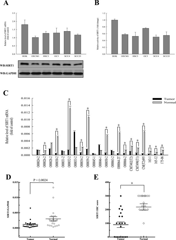
Figure 1.

Variable levels of SIRT1 expression and its activity were noted among normal cells (HOK) and OSCCs. (A) Quantitative RT-PCR (qRT-PCR) and western blotting revealed the expression levels of SIRT1 in HOK and OSCC cell lines. (B) Specific activities of SIRT1 in HOK and OSCC cell lines were determined by enzyme assays. Equal amounts of cellular SIRT1 protein (250 ng) were immunopurified with antibodies against SIRT1, and SIRT1 enzyme activity assays were performed with a SIRT1 Fluorometric Kit, using standard protocols provided by the supplier. (C and D) qRT-PCR revealed significant underexpression (P = 0.0024 ) of SIRT1 in 14 of 21 OSCC samples compared with their matched normal tissues. (E) The expression levels of SIRT1 in the normal and tumor tissues of 21 OSCC patients as determined by IHC. The IHC semi-quantitative score was derived by two independent pathologists who multiplied the staining intensity by the percent of tumor cells stained. IHC scores for each core of a specimen were averaged (n = 21) and statistically analyzed (*, p <0.05). Each data point represents the mean value ± SD obtained from at least three independent experiments.
