Fig 6.
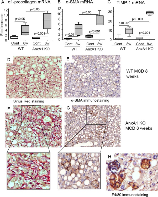
AnxA1 deficiency promotes hepatic fibrosis in mice with NASH. WT and AnxA1 KO C57BL/6 mice were fed the MCD diet for 8 weeks. (A-C) Liver mRNA levels for pro-collagen-1α, α-SMA, and TIMP-1, as measured by RT-PCR, and are expressed as fold increase over control values after normalization to the β-actin gene. Values refer to 6-8 animals per group and boxes include the values within 25th and 75th percentile, whereas horizontal bars represent the medians. The extremities of the vertical bars (10th-90th percentile) comprise 80% of the values. Statistical differences were assessed by one-way ANOVA test with Tukey's correction for multiple comparisons. (D and F) Collagen deposition as detected by Sirius Red staining in representative liver sections from 8-week MCD diet in WT and AnxA1 KO mice. (E and G) Activated HSCs expressing α-SMA (magnification, 400× and 200×). Enlargement shows α-SMA-positive HSCs surrounded by collagen fibers forming cell foci with mononucleated cells. (H) These latter were stained by the macrophage marker, F4/80 (magnification, 600×).

