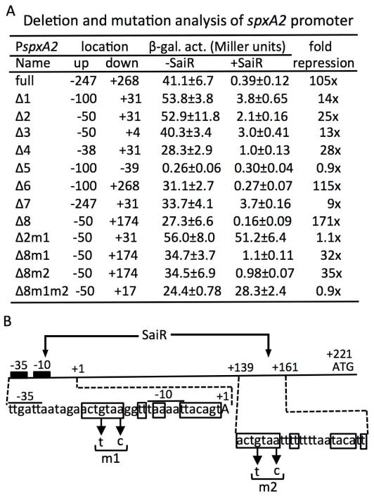
Fig. 4. The spxA2 promoter contains two cis-sites required for SaiR repression.
A. Deletion and mutational analysis of the spxA2 promoter. Endpoints of the promoter fused to lacZ are shown in numbers relative to the transcriptional start site. Each strain was grown in LB and cells were harvested around OD600≈0.4 to measure β-galactosidase activities. Experiments were repeated at least three times using independent isolates obtained from strain construction and the averages are shown with standard deviations.
B. A schematic map of the spxA2 promoter region. The location of the transcription site, the core promoter, and the translation start site are marked as +1, −10 and −35, and ATG, respectively. The nucleotide sequences of the two SaiR sites are shown below the map. Boxed nucleotides constitute a dyad symmetry sequence in site 1 and a partial dyad symmetry sequence in site 2. Arrows show the base substitutions introduced in site 1 (m1) and site 2 (m2).

