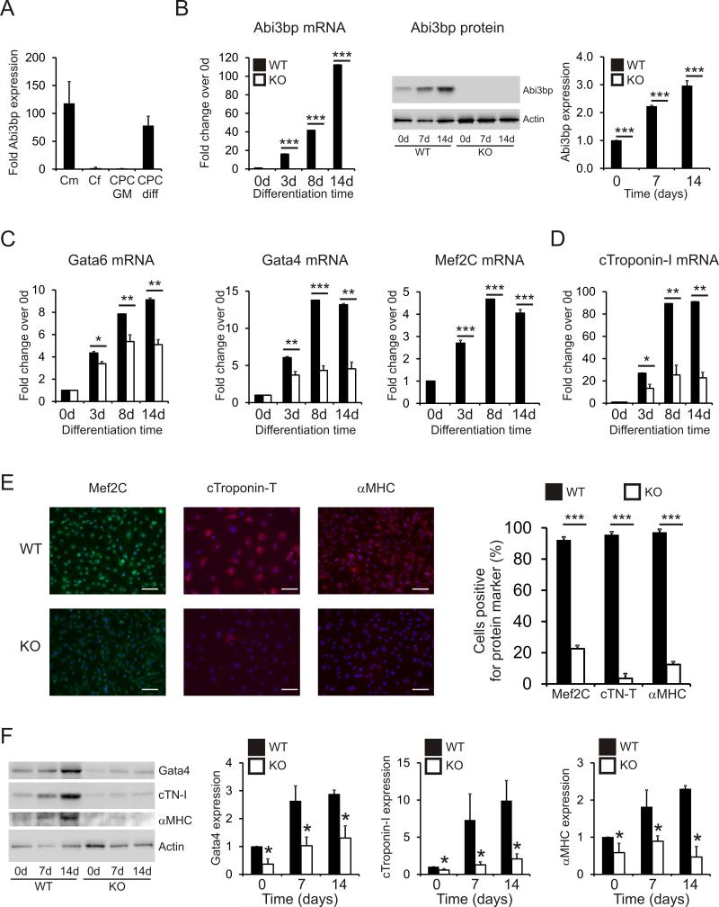
Figure 1. Abi3bp knockout inhibits CPC differentiation.
(A) Abi3bp expression in cardiomyocytes [Cm], cardiac fibroblasts [Cf], and CPCs grown in either growth media [CPC GM] or differentiation media [CPC diff] was determined by qPCR. N=3. Data is shown as a fold expression with c-Kit+ CPCs grown in CPC-maintenance media taken to be 1. (B) Wild-type and Abi3bp knockout CPCs were cultured in CPC-differentiation media for up to 14 days. Expression of Abi3bp was determined by qPCR and immunoblotting. Expression in day 0 wild-type CPCs was taken to be 1. N=3. ***P≤0.001. (C-D) Wild-type and Abi3bp knockout CPCs were cultured in CPC-differentiation media for up to 14 days. Expression of Gata4, Gata6, Mef2C (C), and cardiac troponin-I (D) was determined by qPCR at the indicated time-points. Expression in day 0 wild-type CPCs was taken to be 1. N=3. ***P≤0.001. (E) Wild-type and Abi3bp knockout CPCs were cultured for 14 days in CPC-differentiation media. The cells were subsequently stained with Mef2C, cardiac troponin-T, or αMHC antibodies. DAPI was used to stain nuclei. Scale bar 100 microns. N=4. (F) Protein extracts (7.5μg) from wild-type and Abi3bp knockout CPCs cultured in CPC-differentiation media for 0, 7 and 14 days were probed for the indicated proteins. Actin was used as a loading control. Intensities were normalized to the loading control; normalized intensity of wild-type cells at day 0 was taken to be 1. N=3. *P≤0.05.

