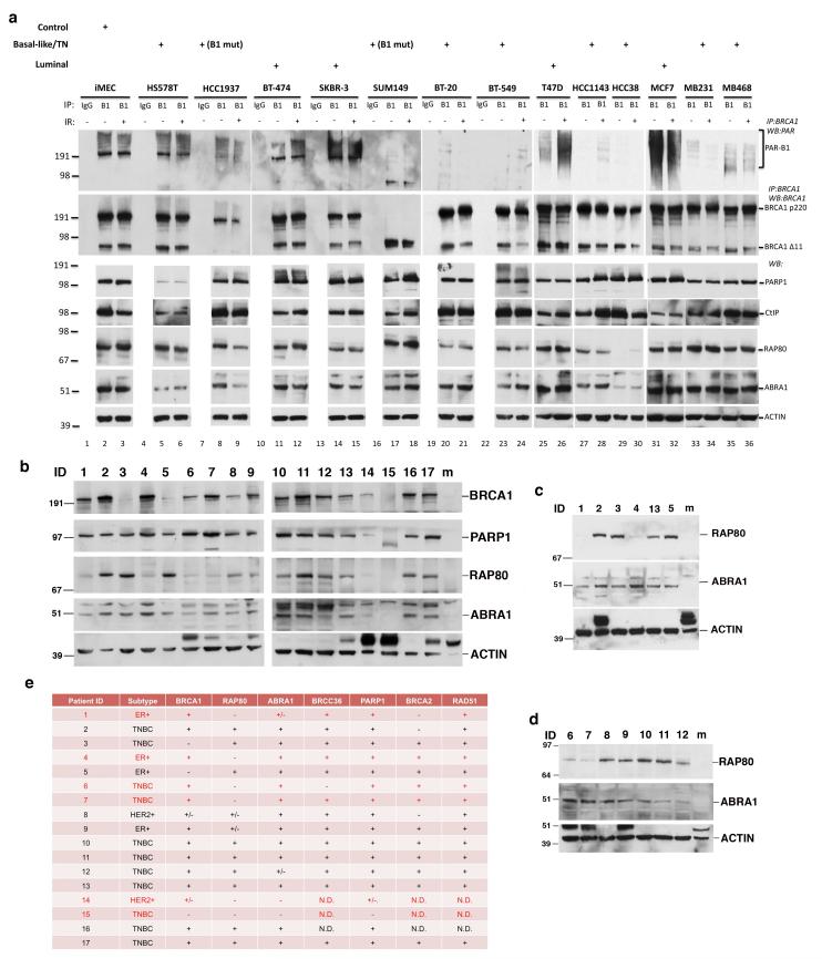
Figure 7. BRCA1 PARsylation and/or RAP80 expression is suppressed in a subset of breast cancer cell lines and tumors.
(a) Results of IP-Western blots searching for BRCA1 PARsylation in a panel of normal breast and breast cancer cell lines (the pathological subtype of each cell line is indicated above the relevant blot; B1-mut: cell lines that contain pathological BRCA1 mutations). Cells were irradiated with 10 Gy IR or mocked treated and collected 8 hours later for analysis. IPs were performed using a rabbit polyclonal anti-BRCA1 antibody. Immunoprecipitated proteins were blotted and probed with a monoclonal anti-PAR antibody (top panels) or a monoclonal anti-BRCA1 antibody (the second row of panels). 20 μg of protein extract from each cell line were blotted and probed with antibodies recognizing PARP1, CtIP, RAP80, ABRA1 and Actin, respectively. (b-d) Results of Western blots detecting BRCA1, PARP1, RAP80, ABRA1 and ACTIN expression in subsets of tumor samples from PDX models. 20 μg of protein extract from each tumor were blotted and probed with antibodies recognizing the above noted proteins. “m” indicates lanes that were loaded with extracts from mouse breast tissues. Please note that mouse Brca1, Rap80 and Abra1 were not detectable by the antibodies used, which specifically recognize human proteins. Results shown were obtained in three, different experiments using independent snap-frozen tumor samples (see Supplementary Methods). (e) Table summarizing the results of protein expression analysis by Western blotting of tumor samples from 17 PDX models. +, positive; −, negative; +/−, weaker expression than other samples; N.D., not determined.

