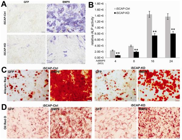
Fig. 4. Silencing β-catenin expression significantly diminishes BMP9-induced osteo/odontoblastic differentiation of iSCAP cells.
(A) and (B) BMP9-induced ALP activity is decreased in the β-catenin silenced iSCAP cells. (A) ALP histochemical staining assay. Subconfluent iSCAP-KD and iSCAP-Ctrl cells were infected with AdBMP9 or AdGFP. At day 5 post infection, cells were fixed for ALP histochemical staining assay. Each assay conditions were done in triplicate. Representative results are shown. (B) Quantitative ALP assay. Subconfluent iSCAP-KD and iSCAP-Ctrl cells were infected with the indicated titers (MOIs) of AdBMP9 or AdGFP. At day 5 post infection, cells were lysed for quantitative ALP activity assays. Each assay conditions were done in triplicate. “**”, p<0.001 (iSCAP-KD vs. iSCAP- Ctrl). (C) Alizarin red S staining. Subconfluent iSCAP-KD or iSCAP-Ctrl cells were infected with AdBMP9 or AdGFP, and maintained in matrix mineralization culture medium for 10 days. Matrix mineralization nodules were stained with Alizarin Red S as described in Methods. Staining results were recorded under a microscope. (D) Oil Red O staining. Subconfluent iSCAP-KD or iSCAP-Ctrl cells were infected with AdBMP9 or AdGFP, and cultured for 10 days. Oil Red O staining was performed as described in Methods. All staining experiments were carried out in duplicate. Representative images are shown.

