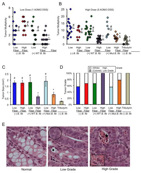
Figure 1.
A high-fiber diet protects against colorectal tumors in a microbiota- and butyrate-dependent manner. (A, B) Scatter plots showing tumor multiplicity for mice in each treatment group following low (A) and high (B) carcinogen exposures. Means are shown, and symbols denote groups with statistically-significant differences: * versus #, p < 0.01; * versus +, p = 0.05; # versus +, p < 0.05. (C) Tumor size presented as mean ± SE with significant differences indicated (* versus #, p < 0.01). (D) Percentage of low-grade versus high-grade tumors for each treatment group based on H&E characteristics of dysplasia within tumors as described in panel E. (E) H&E-stained sections representative of normal colonic epithelium, low-grade tumors, and high-grade tumors. Compared to normal colon, low-grade tumors exhibit hyperplasia (circled region) and are less differentiated with fewer goblet cells that are identified by mucous vacuoles (black dot). High-grade tumors exhibit these features plus a loss of polarity (dotted circled region) and have crypt abscesses with apoptotic material (star). A subset of high-grade tumors display signs of potential invasion based on tumor cells within the muscularis (inset).

