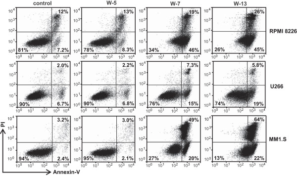
Figure 4.

Effects of CaM antagonists on apoptosis in multiple myeloma cells. Cells were treated with CaM antagonists (60 μM) for 24 h. The cells were subsequently harvested and incubated with FITC-labeled annexin V and propidium iodide and analyzed by flow cytometry. In the representative flow plots, the lower left quadrant contains normal cells; the lower right quadrant contains early apoptotic cells; and the upper right quadrant contains late apoptotic and necrotic cells.
