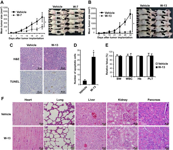
Figure 7.

In vivo antitumor effects of CaM antagonists in a murine multiple myeloma xenograft model. RPMI 8226 cells were implanted subcutaneously into the flanks of nude mice. Seven days later, 3 mg/kg of CaM antagonists were injected intraperitoneally on 5 days in per week. The tumor sizes were measured twice weekly. The comparison groups were the vehicle (H2O) vs. W-7 group (A) and the vehicle (PBS) vs. W-13 group (B). The photographs show representative mice, and arrows indicate the tumors. Tumor tissue sections from the mice treated with vehicle or W-13 were stained with hematoxylin/eosin (H&E) or terminal transferase dUTP nick-end labeling (TUNEL) staining (C). TUNEL-positive apoptotic cells were counted in 10 random high power fields (D). Complete blood count and body weight (BW) were also examined in the mice exposed to vehicle or W-13 (E). Data are from five independent animals and are expressed as the mean ± standard deviation. *P <0.01 compared with vehicle. Histology sections of selected organs from the mice treated with vehicle or W-13 were stained with H&E, and representative tissue section of each organ was shown (F).
