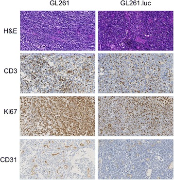
Figure 6.

Histopatholgic and immunohistochemical analysis of tumor specimens from GL261 and GL261.luc bearing animals. Hematoxylin and Eosin (H&E) staining was used for conventional morphologic analysis of tumor. Ki-67 staining was for examining tumor cell proliferation, whereas C3 and CD31 staining was for T-cell infiltration and tumor neovascularization, respectively. Magnification is 200 X.
