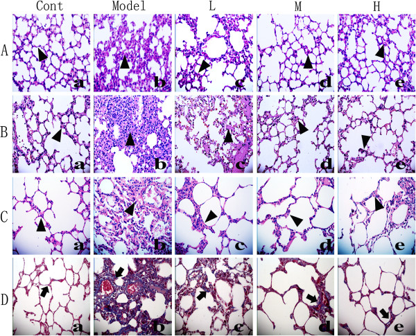
Figure 3.

Effects of NNAV on the histopathological changes in the lung tissue (HE staining and Masson’s trichrome staining, ×400). Animals were treated as described in the legend to Figure 1. Lung tissue was fixed in 10% formalin, sectioned and stained with HE and trichrome. Inflammatory cell infiltration (▲) and the blue color collagen deposition (↑) in the lung tissue were found. HE staining: (A) Six h after LPS. (a) Control. (b) LPS. (c) LPS + NNAV 30 μg/kg. (d) LPS + NNAV 90 μg/kg. (e) LPS + NNAV 270 μg/kg. (B) Eight weeks after LPS. (a) Control. (b) LPS-treated lung. (c) LPS + NNAV 30 μg/kg. (d) LPS + NNAV 90 μg/kg. (e) LPS + NNAV 270 μg/kg. (C) Eight week after BLM. (a) Control. (b) BLM. (c) BLM + NNAV 30 μg/kg. (d) BLM + NNAV 90 μg/kg. (e) BLM + NNAV 270 μg/kg. (D) Masson’s trichrome staining: D(a) Control. D(b) BLM. D(c) BLM + NNAV 30 μg/kg.D(d) BLM + NNAV 90 μg/kg. D(e) BLM + NNAV 270 μg/kg.
