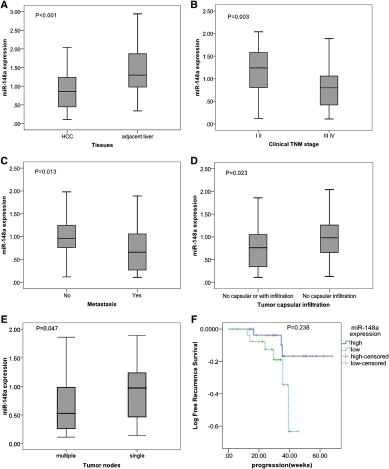
Figure 1.

Clinicopathological impact of miR-148a expression in hepatocellular carcinoma (HCC) tissues. Total miRNA was extracted from HCC and their paired adjacent noncancerous liver tissues. MiR-148a expression was detected by using real time RT-qPCR and the relevant miR-148a level was calculated as compared to the reference of miR-191 and miR-103 combination. Data were shown as mean ± SD. (A) Different liver tissues; (B) Clinical TNM stages; (C) Metastasis; (D) Tumor capsular infiltration; (E) Tumor nodes. (F) The time-to-recurrence of high expression of miR-148a (higher than the median level) was 61.47 ± 3.45 months, longer than those with low expression (50.56 ± 4.15); however, the difference was not significant (P = 0.238).
