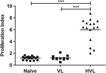
Figure 1.

Lymphoproliferative response to TSLA in VL, HVL and naive groups. Peripheral blood lymphocytes from individuals with active VL (n = 11), HVL (n = 16) and naive (n = 19) groups were incubated with TSLA (10 μg/ml) for 120 hrs and lymphoproliferation was measured by BrdU incorporation for the last 12–14 hrs using BiotrakTM cell proliferation ELISA system. Horizontal lines indicate mean values. ***p < 0.001.
