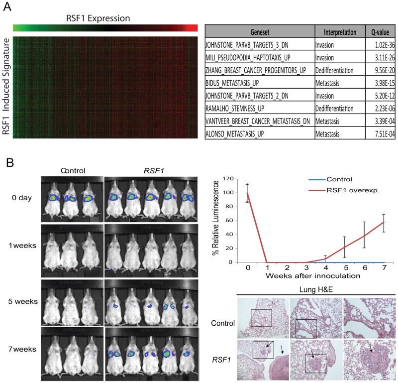
Figure 6. RSF-1 alteration promotes metastasis.
(A) The analysis of the expression changes related to RSF-1 overexpression in basal primary tumors revealed a signature enriched for invasiveness, migration and dedifferentiation (table at right). The heat map at left shows genes in the signature as rows and samples as columns and the color indicates the relative expression (green-low and red-high) and demonstrates the tight correlation of the signature genes across patients. Similar results were observed for luminal primary tumors (Supplementary Figure 6A). See Supplementary Figure 6B-C for analysis of downregulated genes. (B) Comparison of lung metastasis formation in SCID mice subjected to tail vein injection of MCF-10A-TM cells expressing a luciferase reporter and either an RSF-1 over-expression vector or a control vector. H&E of sectioned lungs from mice injected with control and RSF-1 overexpressing cells is also shown. The arrows indicate the presence of metastatic outgrowths in the lungs.

