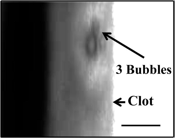
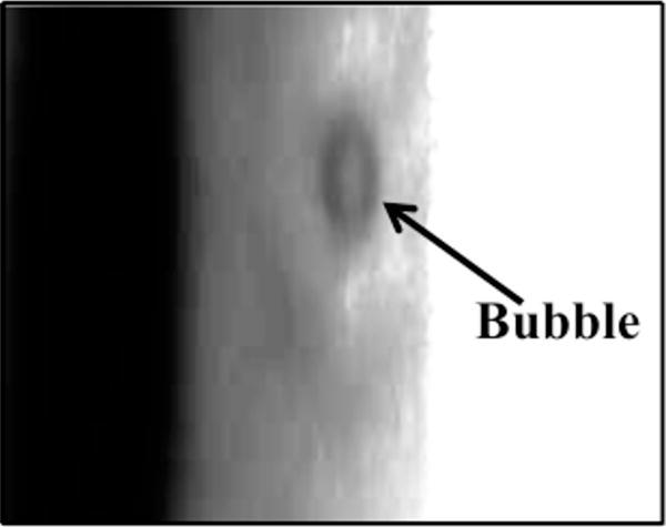
Figure 3.


Coalescence and translation of bubble. (a) Prior to insonation, three bubbles are visible on the clot. (b) The three bubble coalesce within 400 ms after ultrasound exposure. (c) The resultant bubble translates after remaining stationary for 3.5 s. The surrounding fluid contains recombinant tissue-type plasminogen activator (0.32 μg/mL), and Definity® (2 μL/mL). The bubble appears distorted due to the long exposure time of the camera (16 ms) compared to the acoustic period (8.33 μs). The scale bar in panel a is 100 μm.
