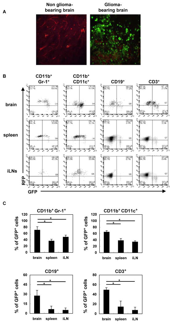
Figure 2. Type-I IFN signaling in the tumor microenvironment.
tdTomato mice bearing SB-induced glioma were sacrificed between days 40 to 50. (A) Brain sections were evaluated by two-photon microscopy for GFP+ and RFP+ cells. Original magnification, 60 X. (B) BILs, splenocytes, and iLN cells were evaluated for the percentages of GFP+ cells. Representative flow histograms are shown. (C) Percentages of GFP+ cells in the glioma-bearing brain, spleen and inguinal LNs (3 mice/group). *p < 0.05 based on Student’s t-test.

