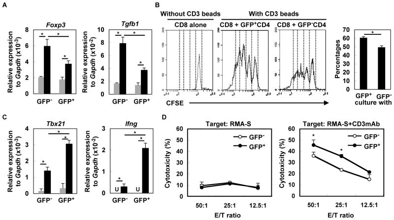
Figure 3. Type-I IFNs directly impact T-cell functions in glioma-developing mice.
(A) CD4+ cells from draining LN derived from glioma-developing tdTomato mice were sorted into GFP− or GFP+ cells and incubated with (black bars) or without (grey bars) anti-CD3mAb. After 4 h, total RNA was extracted for evaluation of Foxp3 and Tgfb1 mRNA levels by qRT-PCR. (B) CFSE-labeled WT CD8+ T-cells were co-cultured with GFP− or GFP+ CD4+ T cells in the presence of CD3 beads. After 60 h, division of CFSE-labeled CD8+ T-cells gated by reactivity to PE-Cy7-condjugated anti-CD8mAb was evaluated by CFSE intensity. As a negative control, CFSE-labeled WT CD8+ T-cells were cultured without any stimulation (left panel). Histograms are representative of two independent experiments. The bar graph shows the percentage of CD8+ cells that have divided at least twice in each of two stimulation conditions (N=4/group; *p < 0.05). (C) GFP− or GFP+ CD8+ T-cells were incubated with (black bar) or without (grey bar) anti-CD3mAb. After 4 h, total RNA was extracted for evaluation of Tbx21 and Ifng mRNA expression levels by qRT-PCR (U: undetected). (D) Cytotoxic activity of GFP− and GFP+ CD8+ T cells was evaluated by 51Cr-release assay. RMA-S cells untreated (left panel) or pretreated (right panel) with anti-CD3mAb (10μg/mL) were used as target cells. Each experiment was performed at least twice. *p < 0.05 compared at the same E/T ratio.

