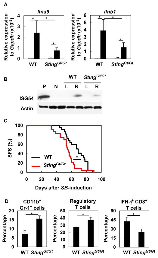
Figure 5. STING contributes to IFN production and anti-glioma immunity.
(A) WT or StingGt/Gt mice bearing SB-induced gliomas were sacrificed between days 40 to 50 (with tumors of similar size at ~1×10⁷ luciferase units). Total RNA was extracted from the left (non-tumor bearing; gray bars) and right (tumor-bearing; black bars) hemispheres of each brain separately (5–6 mice/group), and evaluated for expression of Ifna6 (left) and Ifnb1 (right) by qRT-PCR. *p < 0.05 based on Student’s t-test. (B) Fifty μg protein extracts were analyzed by SDS-PAGE and western blotting for detection of ISG54. “L” and “R” indicate samples from left and right hemispheres, respectively, of WT or StingGt/Gt mice bearing SB-induced glioma. WT-derived macrophage sample treated with p(I): p(C) was used as positive control (P). Glioma-free WT mouse brain was used as negative control (N). Actin was used as an internal control. (C) Gliomas were induced in WT (22 mice; black line) or StingGt/Gt mice (23 mice; red line) neonatal mice by the SB-transposon system. Survival was monitored. *p < 0.05 based on Log-rank test. (D) Percentages of CD11b+ Gr-1+, CD25+ Foxp3+ CD4+ (Treg), and IFNγ-producing CD8+ T BILs were compared between the two groups by flow cytometry. Each experiment except for (C) was performed at least twice. *p < 0.05 based on Student’s t-test.

