FIGURE 4.
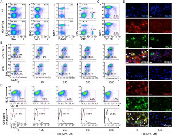
HDI inhibit plasma cell differentiation. (A) Proportions of: B220+PNAhi GC B cells, proliferating B cells (BrdU-stained B220+ B cells), viable (7-AAD−) B220+ B cells and CD4+ and CD8+ T cells in spleen cells from mice that were on HDI-water or untreated water and injected with NP16-CGG 10 d before analysis. (B) HDI does not alter B cell cycle. Mouse IgD+ B cells were stimulated for 48 h with LPS or LPS plus IL-4 in the presence of nil or VPA, during the last 30 min of culture, the cells were pulse-labeled with 10 μM BrdU. The cells were then surface stained for B220 before intracellular staining with anti-BrdU mAb and 7-AAD. B220+ cells are displayed with gates indicating the percentage of cells in G0/G1, S and G2/M phase. (C) Proportions of B220loCD138+ (plasma) cells in spleen cells from C57BL/6 mice that were on HDI-water or untreated water were analyzed 10 d after NP16-CGG injection. (D) Dose-dependent inhibition by VPA of plasma cell (B220loCD138+) differentiation (upper row) in B cells stimulated for 4 d with LPS plus IL-4, without alteration of plasma cell viability, as analyzed by 7-AAD staining (lower row, proportions of 7-AAD− viable cell among B220loCD138+ cells are indicated). (E) IgG1-producing plasma cells (IgG1+CD138+ or IgG1+Blimp-1+) are reduced in cultures of IgD+ B cells stimulated for 7 d with LPS plus IL-4 in the presence of VPA (500 μM), as shown by confocal fluorescence microscopy. Cells were permeabilized and stained with DAPI (blue) to visualize nuclei and fluorescent mAbs to visualize IgG1 (green) and CD138 (red, top set of panels) or Blimp-1 (red, bottom set of panels). Arrows indicate IgG1-producing cells (yellow; CD138+/Blimp-1+IgG1+). Data are representative of three independent experiments; scale bars, 10 μm.

