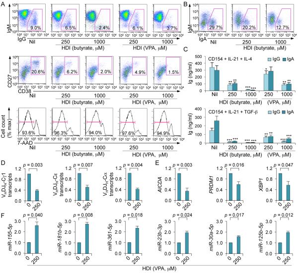
FIGURE 8.
HDI inhibit CSR and plasma cell differentiation increases selected miRNAs, and decreases target AICDA and PRDM1 mRNAs as well as XBP1 mRNA, in human B cells. Human peripheral blood IgD+ B cells were stimulated with CD154, hIL-4 and hIL-21 (for CSR to IgG1 and IgE and plasma cell differentiation) or CD154, hIL-21 and TGF-β (for CSR to IgA) in the presence of nil, VPA or butyrate for 60 h (for transcript or miRNA analysis) or 120 h (for flow cytometry or analysis of Ig titers in supernatants). (A) Proportions of IgG+ B cells, plasma cells (CD27+CD38+) or viable (7-AAD−) CD19+ cells. (B) Proportions of IgA+ B cells. (C) IgG and IgA titers in supernatants of B cells stimulated with CD154, hIL-4 and hIL-21 (top) or CD154, hIL-21 and TGF-β (bottom) and cultured in the presence of VPA or butyrate. Data are from three independent experiments (mean and SEM). *p < 0.05, **p < 0.01, ***p < 0.001, unpaired t-test. (D) Mature VHDJH-Cα (in cells stimulated by CD154, hIL-21 and TGF-β), VHDJH-Cγ1 and VHDJH-Cε (in cells stimulated with CD154, hIL-4 and hIL-21) transcripts were analyzed by qRT-PCR and normalized to HPRT1 transcripts. (E) AICDA, PRDM1 and XBP1 transcripts (in cells stimulated with CD154, hIL-4 and hIL-21) were analyzed by qRT-PCR and normalized to HPRT1 transcripts. (F) miRNA expression was analyzed by qRT-PCR and normalized to expression of small nuclear/nucleolar RNAs RNU6-1/2, SNORD61, SNORD68, and SNORD70. Values in B cells cultured in the presence of HDI are depicted as relative to the expression of each transcript or miRNA in B cells cultured in the absence of HDI, set as 1. Data are presented as mean and SEM from three independent experiments. p values, unpaired t-test.

