
Figure 6.
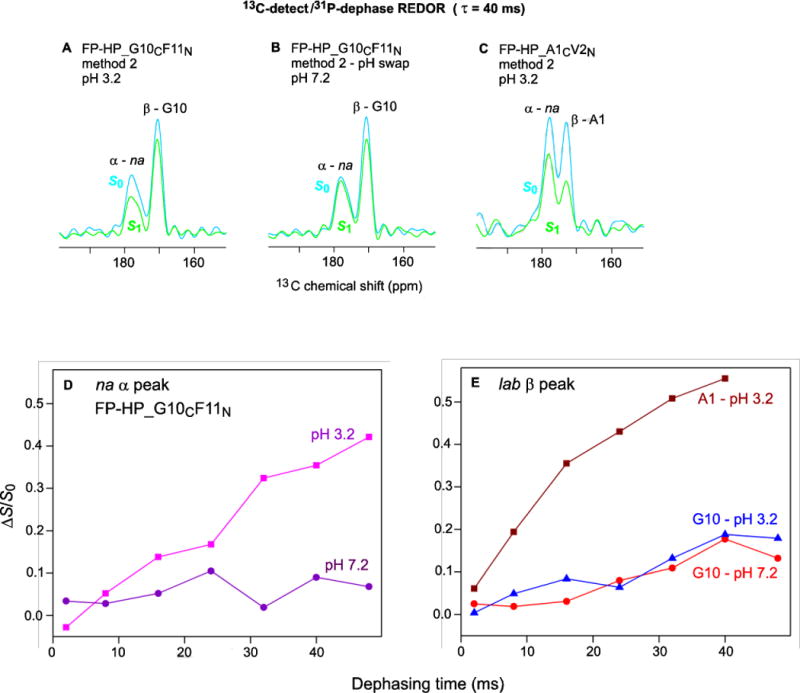
13C-detect/31P-dephase REDOR SSNMR spectra of (A,B) FP-HP_G10CF11N and (C) FP-HP_A1CV2N samples with 40 ms dephasing time. Plots of (ΔS/S0) vs dephasing time for (D) na α signals and (E) lab β signals. Each (ΔS/S0) value was calculated from S0 and S1 13CO intensities integrated over a 3–4 ppm shift interval. The typical uncertainty in a (ΔS/S0) value was 0.01 as calculated from the standard deviation of spectral noise values integrated over comparable interval width.