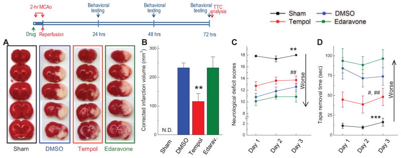
FIG. 3. Neuroprotective properties of tempol and edaravone assessed as drug-dependent reduction in infarction volumes at 72 h and neurological deficits at 24, 48, and 72 h after completion of 2-h MCAo.
In these experiments 5 μL of vehicle (DMSO), tempol (500 nmols), or edaravone (500 nmols) were injected into the lateral ventricle 15 min before initiation of ischemia. Sham-operated animals were subjected to the same surgical procedures without MCAo. Experimental design is depicted in diagram on the top. A, Representative images of 5 serial sections from the same brain (top to bottom) prepared 72 h after reversible MCAo and stained with TTC. B, Average infarction volumes corrected for brain edema in sham operated animals (n=6), and animals injected with vehicle (n=12), tempol (n=11), or edaravone (n=9) as indicated. N.D., infarction not detected. **p<0.01, tempol vs. either DMSO or edaravone. C, Neurobehavioral scores in the same treatment groups, which are presented in B. The score of 18 corresponds to no deficits. **p<0.01, Sham vs. all ischemia groups; #p<0.05, tempol vs. edaravone. D, Average times taken to remove adhesive tape from forepaws. ***p<0.001, Sham vs. all treatment groups; #p<0.05, tempol vs. DMSO; ##p<0.01, tempol vs. edaravone.

