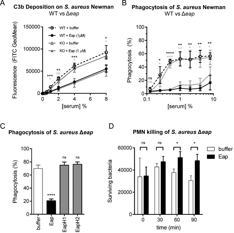
Figure 2. Eap Inhibits Opsonization, Phagocytosis, and Killing of Staphylococcus aureus.
The impact of recombinant Eap on complement deposition and phagocytosis of S. aureus Newman strains was assessed using flow cytometry. (a) C3b deposition on the surface of S. aureus Newman WT or Δeap in the presence of 1 μM Eap or a buffer control. Legend is inset. (b) Phagocytosis of S. aureus Newman WT or Δeap in the presence of 1 μM Eap or a buffer control. Legend is inset in the adjacent panel. (c) Extent of phagocytosis of S. aureus Newman Δeap using 1% (v/v) NHS in the presence of 1 μM Eap, EapH1, or EapH2, or a buffer control. (d) Neutrophil-mediated killing of S. aureus Newman Δeap opsonized in the presence of 1 μM Eap or a buffer control. Error bars represent the mean ± standard deviation of three independent experiments and at least two different donors. Legend is inset. Measures of statistical significance were determined by an unpaired t-test of each experimental series versus the corresponding buffer control for each strain and serum concentration as appropriate. *, p≤0.05; **, p≤0.01; ***, p≤0.001; ****, p≤0.0001; ns, not significant.

